Nagoya City Science Museum
TOP > Exhibition Guide > Keyword Search > Starting with "S" > Scanning electron microscope > Bio Laboratory

Bio Laboratory
Purpose of Exhibition
I would like to try an experiment myself, I would like to try using a microscope…. In response to such desires of visitors, this corner has been transformed from its previous pattern of demonstrating experiments into a corner where all you visitors can experience 20 minutes of experiment and observation using your own head and hands. Here, there are tables and seats where 18 people, in 9 pairs, can sit and conduct experiments. This corner is equipped with biological microscopes, binocular wide-field microscopes, and the scanning electron microscope that is indispensable for micro level observation of organisms. In addition to the biological experiments carried out every day, we will also hold special events and observation sessions using the scanning electron microscope. We have all the tools and reagents necessary for the experiment ready for you, so you do not need to bring anything with you in order to participate. There is no need to register ahead of time. This corner is first come, first served. Please try taking on the challenge of experiment and observation in the life sciences. (For details on the themes of the experiments and the times, please check in the visitor's guide or the Science Museum homepage.)

Additional Knowledge
The development of biology has been supported by the development of observation tools and analytical equipment and by the selection and development of model organisms. Here, we will introduce the scanning electron microscope, which is also found in the Bio Laboratory.
The smallest that we can see with our own eyes is about 0.2 mm. Observing organisms smaller than that or cell forms requires the tool called the microscope. Since the father and son Janssen combined glass lenses to invent the microscope in Holland at the end of the 16th century, the structure of microscope, lens, methods to make preparations etc. have been improved. However, the limit on the smallest size that can be seen with an optical microscope is about 0.2 µm. In the 1930s, the electron microscope, which uses electron beams instead of light for observation, was invented. Electron microscopes include transmission electron microscopes (TEM), which observe thin specimens a few tens of nm thick, and scanning electron microscopes (SEM), which observe the surface of specimens.
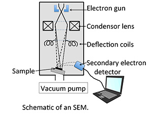
In a scanning electron microscope, the electron beam focused by the lens which comprises magnets, not glass is shone on the specimen and the secondary electrons coming from the specimen are received by the detector and converted into electron signals, which are displayed on the monitor (nowadays, a computer screen). The electron beam is not shone on the specimen as a whole, but rather fine electron beams trace along the specimen surface to obtain the overall image. That is why this is called a "Scanning" electron microscope. It is appropriate for viewing the fine surface structure of a specimen. However, it cannot see colors. Compared to transmission electron microscopes, the preparation of specimens for observation is comparatively simple. Specimen pieces are cut small, fastened using chemicals, then dried. Then a fine layer of heavy metal is vapor deposited on the surface of the specimen and that specimen is observed with the electron microscope.
Recently, the fine structures of organisms have been drawing attention from the perspective of technology. For example, adhesive tape made with mimicking the foot's sole of the gecko at nano -size level seems to be developed.The world that a scanning electron microscope shows is very interesting.
Article by Chieko Ozaka,curator