Nagoya City Science Museum
TOP > Exhibition Guide > Keyword Search > Starting with "F" > fetus > Fetal Growth and Development

Fetal Growth and Development
Purpose of Exhibition
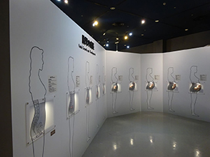
A baby resides in the mother’s womb for about 38 weeks. This exhibit divides this period into 10 segments and explains them with models, explaining the baby’s growth process and describing the characteristics of the individual segments.
A baby residing in the mother’s womb is called a “fetus.” The body system of a fetus develops at an incredibly high speed.
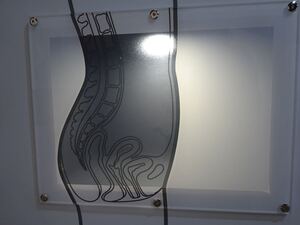
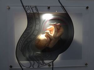
Each model shows a longitudinal section of a mother’s belly, illustrating how it swells, as well as how the baby grows within it. Note that these models were originally installed in the Life Science Building when it was opened in the Nagoya City Science Museum in 1989.
[Differences from the Original Exhibit]
Descriptive text about the individual segments has been modified in reference to the materials including those currently used by Nagoya City. In addition to the pregnancy week count, the number of weeks after fertilization is also mentioned. Illustration of a placenta have been newly added. The illustrations of the sagittal section of the mother’s body and the womb have been remade based on previous exhibits, whose figures were copied down. Dr. Schweitzer's words, which used to be displayed near the "Fertilization and Delivery", the exhibit before the remodeling, have been displayed again.
Additional Knowledge
Related information is given with other exhibits and descriptive text in the “Growth, Reproduction and Aging” zone.
[The Pregnancy Week Count and the Number of Weeks After Fertilization]
“Week XX of pregnancy” differs from “week XX after fertilization.” Fertilization does not produce any symptoms that the mother is conscious of, and not all fertilized eggs are implanted. So, using the fact that pregnancy stops the menstrual cycle, the first day of the menstrual period before fertilization (called the “last menstrual period”) is counted as day 0 of week 0 as the base point in time. Usually, menstruation starts about two weeks before ovulation. So based on this counting system, weeks 0 to 1 of pregnancy are, in most cases, before ovulation, which means that, in a strict sense, the mother is not yet pregnant.
Today, at elementary school, pregnancy is taught in science class and health and physical education class based on the number of weeks after fertilization. It may be difficult for visitors to understand pregnancy with only the pregnancy week count. So, this exhibit shows both of these counting systems.
[Placenta]
When a fertilized egg is implanted and keeps dividing, a placenter is formed. The placenta is a structure that supplies nutrients and oxgen from the mother's body to the fetus and passes waste products and carbon dioxide to the mother's body.
The placenta is formed at the end of the umbilical cord jointly by the fetus (the fetus in the early stages is called an “embryo”) and the mother. Some cells of the fetus divide to form membranes which develops branches like a tree to have many thorn-like projections, inside which blood vessels from the umbilical cord run. These thorns are wrapped by membranes formed by the mother’s body. Between these two membranes, blood from the mother’s body flows to exchange gas and other substances. The placenta has several other functions, including producing a hormone that plays a key role in maintaining pregnancy.
[Growth]
A fertilized egg measures about 0.1 mm in diameter and can barely be seen with the naked eye. Growing in the womb, the baby measures about 50 cm in height and weighs about 3,000 g by the time of birth.*
* The height and weight vary from baby to baby.
Article by Tomoko Horiuchi, curator