Nagoya City Science Museum
TOP > Exhibition Guide > Keyword Search > Starting with "N" > nerve > How Your Body Works

How Your Body Works
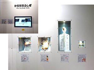
Purpose of Exhibition
The screen plays a video with a narration, which tells you that changes continue to occur all over your body without your awareness of it, and that a human body is filled with wonders.
Around the screen, various model organs are exhibited with descriptive text. Through them, you can learn how internal organs work to play their various roles.
Note: This exhibit reuses the model organs originally installed at the opening of the Life Science Building in the Nagoya City Science Museum in 1989, and describes digestive tubes, the liver, the central nervous system, and teeth with these exhibits.
[Differences from the Original Exhibit]
The following items have been remade: a diagram illustrating the human body is displayed at the back, and nameplates of individual body parts are in the front.
Additional Knowledge
Related information is given with exhibits and descriptive text in the zones “Nervous System and Endocrine System” and “Digestion, Absorption and Excretion.”
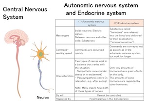
[Homeostasis to Keep the Inside of the Body Balanced]
The video explains how various parts of the body act together to work well—without our awareness of it—to maintain a similar condition. This system is called “homeostasis.” When the condition of the inside or outside the body changes, that change is conveyed by sensory nerves to the central nervous system (the brain and the spinal cord), and the autonomic nervous system and the endocrine system interact with each other to keep the body in the right condition.
For example, when it is hot, the body works as follows to prevent the body temperature from rising too high. Muscles and the liver break down fewer substances to produce less heat. Vessels expand to release more heat from the skin. Sweating, too, prevents an excessive increase in body temperature. When the sweat evaporates, heat is conducted away from the skin surface (“heat of vaporization”).
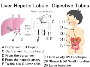
[How Does Food Change in Digestive Tubes?]
When you take in food through your mouth, nutrients are taken into your body in the order listed below. In addition to the long digestive tubes, which are connected to each other, the digestive organs, including the pancreas, the liver and the gallbladder, deliver digestive juice to extract nutrients from the food.
(1) Oral cavity: The food is chewed with the teeth, and starch found in the food is digested to some extent. Then, the food is sent to the esophagus.*1
(2) Esophagus: Sends the food to the stomach in small amounts
(3) Stomach: Absorbs alcohol, carbonic acid, and certain types of medicine, and breaks down protein
The stomach is shaped like the letter “J,”*2 and sends food to the intestines.
(4) Small intestine: Breaks down protein, starch and fat with pancreatic juice and intestinal juice (a digestive enzyme from the wall of the small intestine) to absorb the nutrients, which are carried by the capillary vessels around it (fat is carried by lymphatic vessels).
(5) Large intestine: Loops around the belly like an upside-down U and absorbs water.
*1 When the food is sent to the esophagus, “flaps” work to prevent the food from entering the windpipe and the nasal cavity.
*2 Its inclination and curve vary from person to person.
[Liver and Gallbladder]
The model liver displayed shows how large and massive this organ, one of the largest in the human body, actually is. The model gallbladder displayed shows what this sack-shaped organ looks like, which stores bile, a digestive juice that is important in digesting and absorbing fat.
The “Fine Structure of the Liver” section displays a model of the liver’s basic component called a “hepatic lobule,” (a polygonal prism that measures about 1 mm in diameter and about 2 mm in height, with a blood vessel running through the center). With a closer look at the model, you can recognize elements of the complex structure of the liver: the portal vein*4 (A) sends blood from the digestive tubes to the liver cells; the hepatic artery (B) sends oxygen-rich blood to the liver; thin blood vessels (D and E) branch off from the portal vein and the hepatic artery respectively; thin blood vessels (C) lead to the hepatic vein, which carries blood from the liver; and tubes (F) lead to the bile duct, which sends bile to the gallbladder to collect the juice there. (G) indicates the liver cells.
[Is the Liver a Chemical Plant?]
The liver can be compared to a chemical plant because many chemical reactions take place in this organ. The liver plays the following three major roles:
(1) Storage, synthesis and breakdown
• To synthesize (make) glycogen from glucose*1
• To synthesize and break down many types of protein
(2) Detoxification
• To neutralize substances that are harmful to the human body
E.g., to break down alcohol into acetic acid in the end*2 and to turn ammonia into urea
(3) Production of bile (a digestive juice important for fat digestion and absorption)*3
*1 When the glucose level in the blood decreases, glycogen is broken down into glucose.
*2 Acetaldehyde, a highly toxic substance, is formed midway in the process.
*3 Bile is stored in the gallbladder.
*4 A blood vessel that has capillary vessels on both ends is called a “portal vein.” The one for the liver is a typical portal vein and is also called the “hepatic portal vein.”
[Central Nervous System]
Nervous systems enable you/your body to learn what is going on inside and outside the body and to react to the situations. The systems mainly consist of special cells called “nerve cells” (also called “neurons”), which are scattered throughout the body.
More complex larger nervous systems with more neurons convey more information. The central nervous system serves as a control tower in which information is concentrated and from which information is conveyed as appropriate.
Vertebrates, or animals with a spine, have a central nervous system developed along the back into a thick cord called the “spinal cord.”
The top of the spinal cord has swollen into an especially developed organ called the “brain.” The brain consists of the cerebrum, the midbrain, the cerebellum, and the diencephalon.
Between the brain and the spinal cord is a swelling called the “medulla oblongata” which serves as the mainstay of breathing and reflex.
This model shows an overview of the central nervous system, which supervises the nervous systems in the human body. As you can clearly see, the human cerebrum has developed so large that it almost covers the rest of the brain, which is invisible from the outside except for some parts of the cerebellum and the medulla oblongata.
[Are the Mouth and Teeth Only for Food?]
The oral cavity is a place that holds the lips, cheeks and jaws together and is connected to the throat at the back. The oral cavity is the entrance for food and air, and is deeply connected to your daily life. You can taste food with the tongue, cram a lot of food into your mouth thanks to the right and left cheeks, chew the food with your teeth, swallow it, and pronounce words.
When the vocal cords in the airway vibrate to form sounds, the oral cavity adds various resonance and tonal components to produce words and voice, which is another important role of the oral cavity.*
* The nasal cavity and the skull, too, are involved in pronunciation.
Article by Tomoko Horiuchi, curator