Nagoya City Science Museum
TOP > Exhibition Guide > Keyword Search > Starting with "S" > synapse > Test Your Own Reaction Time

Test Your Own Reaction Time

Purpose of Exhibition
Put your finger on the Experiment Switch and remove it as soon as you notice a signal. A signal is either a light or sound. Your reaction time, time between the moment the signal is emitted and the moment you remove your finger from the switch, is measured three times for each type of signal. Your average reaction time is also displayed.
Try this experiment to see to which signal you react more quickly. If you are here with your friends, it would be interesting to compare the results. Please note, however, that only one person can experiment at a time.
Additional Knowledge
You see and hear because your eyes and ears convey information to the brain. Let’s see how this process works with illustrations.
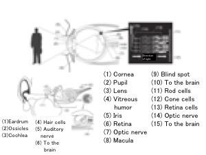
[Mechanism of seeing]
Vision – Light enters the eye and is reflected on the retina inside the eye, and the optic nerve conveys information, such as the intensity and color of light rays, in the order described below.
Cornea is the transparent film protecting the pupil and the iris of the eye
Pupil is the dark center of the eye. The pupil looks dark because the light that enters the eye is not reflected, but it does not have a dark color. If you look at your pupil closely in the mirror, you can tell that it becomes larger or smaller, depending on how much light there is around you.
Iris is the colored part around the pupil. It changes the size of the pupil to regulate the amount of light that enters the retina. The color of the iris varies from one person to another, depending on the amount of melanin pigment. The majority of Japanese have brown irises.
Lens in the human eye can flexibly change its thickness to adjust focus, according to the distance between the eye and the object. When you look at an object near you, the ciliary body around the lens contracts, relaxing in turn ciliary zonule which pull the lens outward, thereby thickening the lens and allowing it to focus on the object.
The lens can become less flexible with age, leading to presbyopia (difficulty in clearly seeing objects nearby).
Retina is at the back of the eye. Light that has passed through the vitreous humor behind the lens reaches the retina. Two types of cells that sense light (visual cells) are found here side by side:
- Cone cells function in bright light and sense colors and shapes clearly.
- Rod cells function in dim light and do not sense colors and shapes clearly.
Information (stimuli of light) received by the optic cells is sent to the bipolar cells, and from the optic nerve to the visual cortex of the brain.
Macula and blind spot
Optic cells, especially cone cells, are concentrated in the macula, near the center of the retina. Images reflected here are particularly clearly seen.
The optic nerve and blood vessels enter and leave the retina at the blind spot, near the nose. Since optic cells are absent here, images reflected here cannot be sensed.
[Mechanism of hearing]
Structurally, the ear comprises the outer ear, middle ear, and inner ear. Auditory information, such as the intensity and height (vibration) of a sound outside the ear, are conveyed in the steps described below:
Outer ear: This is the part from the externally visible part of the ear (pinna) to the eardrum. It is shaped in such a way as to receive sound.
- Eardrum: A thin tissue inside the ear at the end of the outer ear; sound (waves of air) vibrates the eardrum.
Middle ear: This is the part between the eardrum and the inner ear. The Eustachian tube leads to the throat.
- Ossicles: Three small bones called ossicles are attached to the middle area. Vibration is conveyed to them one after another.
The ossicles are the smallest bones in the human body and are small enough to be placed on a fingertip. From the eardrum to the inner ear, they are called (a) malleus, (b) incus, and (c) stapes, which mean (a) hammer, (b) anvil, and (c) stirrup.
Inner ear: This is the part farthest from the outside. The cochlea, which looks like a snail, is found here.
- Cochlea: Inside this snail-shaped organ, cells and nerves perceive sound. The cochlea is filled with a lymphatic fluid.
The spiral of the cochlea has a three-story structure. Sound vibrations is first conveyed to the top story, to the very end, returning to the bottom story to let the middle story floor vibrate. Depending on the height of the sound, vibration is particularly strong at a specific place (high note near the entrance, low note near the end).
- Hair cells, cells with sensory hair, in the inner ear vibrate to convey sound signals.
- Auditory nerve interprets the vibrations of hair cells and conveys information on the sound (loudness, height, etc.) to the brain.
- The brain receives information about sound.
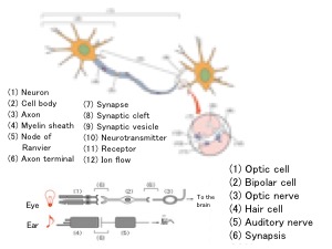
[Synapse, joint of nerve cells]
Inside a nerve cell (neuron) that has received a stimulus, an electric signal is conducted from the cell body to the axon end. In human neurons, myelin sheath that covers the axon and the node of Ranvier increases the conduction speed.
Information from a stimulus is conveyed to the brain not always by a single cell. Information is generally conveyed by two or more cells that relay the signal by turns. Two nerve cells are not connected. There is a very narrow space between them (called synaptic cleft). The structure including the cell membrane before and after the synaptic cleft is called synapse.
[Transmitting information across the cleft]
Neurotransmitters are released from the synaptic vesicle at the end of the axon (axon terminal) of the neuron. Signals are sent when the neurotransmitters are bound with receptors of another cell (nerve or muscle). Ion flows into cells are also involved in this process. Since there are more neurons and synapses involved in the process of vision than in the process of hearing, the reaction time is generally believed to be longer for vision.
The exhibit “Wonders of Nervous System and Endocrine System” introduces the mechanisms of neurons and synapses.
Article by Tomoko Horiuchi, curator