Nagoya City Science Museum
TOP > Exhibition Guide > Floor Map> Gallery of Internal Organs

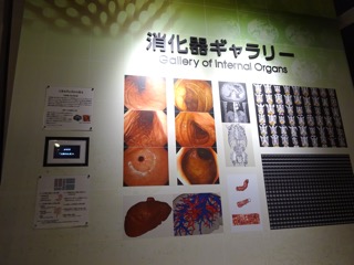
Gallery of Internal Organs


Purpose of Exhibition
This is a corner where you can learn about the sophistication and wonder of organs and tissues through images involving digestion, absorption, and excretion.
The exhibit contains 14 large wall-mounted images along with two surrounding explanations, and images and video displayed in digital photo frames.
Additional Knowledge
Let's look at one of the large images
The gastrointestinal tract seen through an endoscope
Endoscopes can take a picture of the inside by inserting thin tubes into the gastrointestinal tract from the mouth, nose, or anus.
They can inspect things without harming the body, and the tubes can incorporate surgical instruments to perform examinations and surgery at the same time.
In recent developments, the wavelength of light is used to make it easier to find lesions, and a capsule camera has been created that wirelessly transmits images to a device outside the body.
Image (1)-(3): Stomach membrane taken with endoscope
Photograph of the stomach taken with an endoscope. You may be familiar with the 'stomach camera' inserted in the mouth or nose during a health examination.
Image (4)-(6): Mucous membrane of the colon taken by endoscope
Here is a picture of the large intestine taken with an endoscope inserted in the anus during an examination.
[Looking at the body from the outside]
A CT scan can display a cross section of the body by using a computer to process X-ray images taken from different angles.
The X-ray penetrates differently depending on the type of atom, and the difference is expressed as hardness in the image.
This exhibit displays CT images with 0.5mm intervals processed into a virtual three-dimensional human body.
*CT: Computed Tomography
Image (7)-(9): CT image near the kidneys and virtualized 3D shape
You can see that there are many complex blood vessels around the kidneys that regulate the concentration of substances in the body. The inside of the kidney is packed with even finer structures.
Image (10): The size, shape and position of organs varies between people
The images of many people's bodies have been processed to make the organs clearer. Just as each person has a different face, the size, shape, and position of the organs are also different. Your body is one of a kind. Your precious body is yours alone.
Image (11)-(12): An entire virtualized liver and internal blood vessels
Similarly, you can see the fine blood vessels in the liver which works in complex ways.
Image (13): The large intestine is virtually cut open in a planar shape. Image processing has a wide range of applications. The curved shape of the intestines is easy to see, which makes polyps easier to find.
Image (14): Continuous CT image of the chest (top, bottom, left, and right of image correspond to the abdomen, back, right, and left)
A series of CT images ("circular slices" of the body) near the chest of a person. The continuous cross sections allow you to see different organs and their positions.
Courtesy
(1)-(6): Departments of Gastroenterology and Metabolism,
Nagoya City University Graduate School of Medical Sciences
(7)-(9),(11)-(14): Kensaku Mori Laboratory, Nagoya University
(10): Kensaku Mori Laboratory, Nagoya University, "Computational Anatomy" and "Multidisciplinary Computational Anatomy" Grant-in-Aid for Scientific Research on Innovative Areas, MEXT, Japan
Article by Tomoko Horiuchi, curator