Nagoya City Science Museum
TOP > Exhibition Guide > Keyword Search > Starting with "L" > lymph > Wonders of Respiration and Circulation

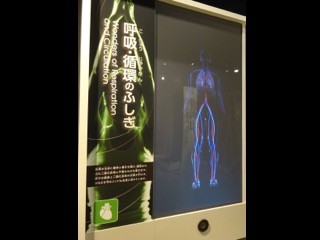
Wonders of Respiration and Circulation
Purpose of Exhibition
This exhibit presents the basics about of respiration and circulation (blood, which delivers oxygen and nutrients to the body, transports carbon dioxide and unnecessary substances from cells; the heart, which delivers a constant supply of blood; the lungs, which exchange oxygen and carbon dioxide; and lymph, which flows throughout the body to protect it).
They are introduced through movie and three pieces of information about each using text and drawings on the reverse side.
Additional Knowledge
The heart is made up of four characteristic chambers. Now, let us introduce you about them briefly. The "left" and "right" referred to herein mean the "left" and "right" in the gigantic human body replica.
(1) Left Atrium (upper left chamber) The left atrium is where oxygen-rich blood arrives from the lungs, and it leads to (2). Attached to the inlet of (2) is a valve that prevents blood's backflow.
(2) Left Ventricle (lower left chamber) The left ventricle pumps oxygen-rich blood to all parts of the body. For this vital function, it is most muscular of all the chambers and its wall is thick. (For its thickness, check it in the exhibit "Heart.") Immediately after passing through the left ventricle, blood is divided into two flows upward and downward flows.
(3) Right Atrium (upper right chamber) The right atrium is where blood returns from all parts of the body and leads to (4), and, of course, there is a valve between (3) and (4).
(4) Right Ventricle (lower right chamber) The right ventricle pumps blood out and into the lungs. In the exhibit "Heart," you will find the wall of the right ventricle is much smaller in thickness than that of (2). Think about the reason why it is so.
[Blood Circulation]
You can now understand that blood is circulated repeatedly in the same order of (1) ― (2) ― entire body ― (3) ― (4) ― lungs ― (1). In this process of blood circulation, the segment of (4) ― lungs ― (1) is called "pulmonary circulation" and that of (2) ― entire body ― (3) is called "systemic circulation".
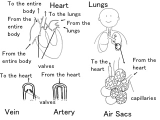
[Lungs]
Air inhaled through the mouth and nose passes through the throat into the trachea, which divides into the left and right bronchi. In the lungs, the airways divide again and again, becoming narrower and narrower, thus ending up as grape cluster-shaped thin bags called "lung's air sacs," which are surrounded by capillaries in a net-like form. The exchange of oxygen and carbon dioxide takes place through the two thin walls of the lung's air sacs and capillaries.
[Artery, Vein and Capillary]
The blood vessels that carry blood away from the heart are called "arteries," and the blood vessels that carry blood back to the heart are called "veins".
The two types of blood vessels are different in wall thickness. You may wonder why arteries have a thicker wall than veins. This can be explained by the fact that blood still has momentum in the arteries, which are closer to the heart; therefore, their wall must be thick enough to prevent wear and tear and to be surrounded by muscles to pump blood. On the other hand, once blood travels around the body, it no longer has momentum when it flows through the veins. So the veins' wall does not need to be as thick as the arteries'. Instead, valves are attached to the veins to prevent momentum-free blood from flowing backward. Those valves open in only one direction, namely, toward the heart, like single swing doors. Should blood happen to flow backward, the valves are closed and no backflow occurs.
The farther away from the heart arteries and veins are located, the thinner they become, and these two types of blood vessels are connected with the netting of capillaries, each of which is thinner than a human hair. Although very thin, the capillaries perform a vital networking function to deliver oxygen and nutrients to each cell of the body and pick up carbon dioxide and body wastes.
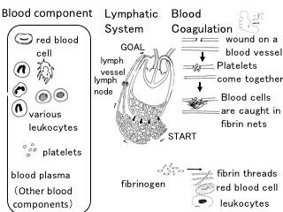
[Blood components and their role]
Blood contains red blood cells (that carry oxygen), white blood cells (that protect the body), and platelets (that clot wounds), which can be seen as 'grains' when viewed with a microscope. There are different types of white blood cells classified by structure and function.
[Lymphatic System]
Lymph (which is also called "lymph fluid") is a clear fluid of plasma that leaks out of the capillaries to surround and bathe the body tissues, and contains more water and less protein than blood and neither red blood cells nor platelets at all. Instead, it contains plenty of lymph cells white blood cells of a kind.
The exclusive vessels through which lymph flows are called "lymph vessels". Along the lymph vessels in the body are lymph nodes, which serve as lymph filters. The lymph near the intestines contains a large amount of intestine-absorbed fat, which is called "chyle".
Lymph is carried through the lymph vessels to the base of the neck where it merges into the bloodstream in the veins and then into the heart.
[Lymph Vessels and Lymph Nodes]
With the arteries and the veins connected with the capillaries, the vascular system is existent in the form of a loop with neither its starting point nor its ending point. But lymph vessels have their origins. The lymphatic system originates with the closed ends of the lymph vessels located at the tips of the hands and the feet. Located along the lymph vessels toward the base of the neck are lymph nodes, which function to remove bacteria, foreign substances, toxins, etc. When you sustain an injury or fall ill, you may have a pain in one of your lymph nodes. If you touch that part, you may feel a lump, which is the swollen lymph node resulted from a bacterial infection or something.
Incidentally, the lymph nodes are where lymph cells multiply.
[Blood Coagulation]
Blood contains a wound-sealing ingredient to prevent a wound from serious bleeding and also protect it from foreign substances.
First of all, platelets in the blood form blood clots to seal the wound. Subsequently, the blood clots are reinforced by red blood cells and white blood cells, which are twined in the net created by intertwined "fibrin" proteins. This first-aid treatment keeps blood from flowing in the blood vessel. Therefore, when the wound gets out of emergency, a substance called "plasmin" dissolves the fibrin so that blood can resume flowing.
[When Fibrin is Formed]
Fibrin's constant presence in the blood blocks the flow of blood. To avoid it, a water-soluble protein called "fibrinogen," which is fibrin's primary form, is normally present in the blood. When part of a blood vessel is wounded, a chemical reaction occurs to transform the fibrinogen into a long chain of the fibrin, which coagulates blood to seal the wound. More than 15 substances are complexly involved in these bio-chemical reactions, and the interaction among the substances has not yet been raveled completely.
Article and illustrations by Tomoko Horiuchi, curator