Nagoya City Science Museum
TOP > Exhibition Guide > Keyword Search > Starting with "B" > bone marrow > Wonders of Muscles / Wonders of Bones

Wonders of Muscles / Wonders of Bones
Purpose of Exhibition
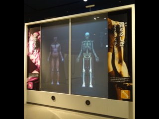
This exhibit presents the basics about muscles (how they contract (shrink) to create the power that allows you to move your body as you intend, and operate your heart, digestive tract, and other organs without any conscious effort), and bones (how they support the body, protect it, make blood, and retain calcium).
Muscles are presented on the left half of the screen, with bones on the right half, and three facts about each using text and illustration on the reverse side.
Additional Knowledge
[Structure and Function of Skeletal Muscle]
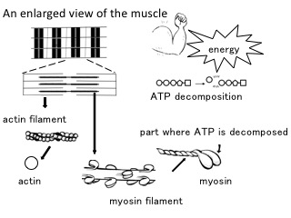
Skeletal muscle is composed of a bundle of long and thin cells, each of which is about 0.02 mm in thickness. This cell is called "muscular cell" or "muscle fiber." Each muscle fiber (muscular cell) is a bundle of strands, each of which is known as "myofibril" and is about 0.001 mm in thickness. Muscular contraction is due to myofibril's contraction, which takes place in the microscopic world.
Myofibril has two different kinds of thin filaments (actin and myosin) aligned in parallel. When actin filaments slide into myosin filaments, myofibril's contraction occurs.
The decomposition of a substance called "adenosine triphosphate (ATP) is the immediate source of energy for muscle contraction. And muscle contraction is characterized by a highly efficient use of the energy with a low rate of thermal loss. It is known that each myosin filament's double-headed portion has the function of converting chemical energy in the form of ATP into mechanical energy. But there remain multiple unanswered questions about how actin filaments are made to slide into myosin filaments through the use of the energy.
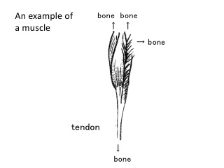
[Tendons transmit skeletal muscle movement to bones]
The muscles cannot push or stretch themselves. They can only contract and relax. So when you need to move your joints in different directions, you need to have different kinds of muscles.
With plenty of strong fibers, the ends of our muscles take a form of glittering white tendons, which are firmly connected with the periosteum that covers bones.
For this strong connection, the contracting force of muscles is transferred to bones. Therefore a bone can act like a lever.
The largest tendon in the human body is the Achilles tendon below the calf muscle.
[Bones and cartilages]
This exhibit shows you that approximately 200 bones make up the adult human body. They are the hard parts of the body. There are also soft and elastic cartilages in ears, the nose, and at the end of and in between bones.
[Structure of Bone]
Bone is not anything like a rigid "stick". Surprisingly, bone is porous. So this particular physical property makes bone light and not prone to fractures. In the pores lie blood vessels and soft bone marrow where blood's ingredients are produced. When you have a food with bone, look at the cross-section of the bone to check its bone marrow.
New bone is constantly being made while old bone is destroyed. During exercise, the function of “osteoclasts” that break down bone is suppressed, while the function of “osteoblasts” that build bone is activated, making bone strong.
[Joints]
The connection between two bones is called a joint.
Numerous ligaments serve to firmly unite the bones to fix the joints securely.
A joint moves when the muscles attached to the bones that make up the joint contract.
Article and illustrations by Tomoko Horiuchi, curator